Diagnosis Using Magnetic Resonance Imaging
")
Magnetic resonance imaging is a diagnostic method based on obtaining “slices” of the area being examined. This method allows for high-quality visualization of various organs and body parts. MRI is based on the phenomenon of nuclear magnetic resonance. To explain the principle of operation in simple terms, an MRI machine is a large magnet that affects the entire human body, causing a change in the response of atomic nuclei. In other words, the nucleus of a hydrogen atom changes. These changes are recorded by the device and converted into an image using special algorithms. Simply put, the method is based on the “saturation” levels of various tissues and organs.
Article contents:
Safety
Originally, the method was called nuclear magnetic resonance imaging. However, following the Chernobyl nuclear power plant accident, any mention of its “nuclear” origin caused some concern among the public. Therefore, it was decided to drop this term in favor of magnetic resonance imaging. In fact, unlike X-rays or CT scans, MRI does not expose the body to radiation. A magnetic field is extremely safe. It surrounds us everywhere. Cell phones, microwave ovens, televisions, and other household appliances are sources of magnetic fields, but with lower “intensity.”
The impact of MRI on human health has been studied since the method was introduced into medicine: over more than 40 years, scientists have not been able to identify any negative effects where the examination posed a specific risk. Therefore, magnetic resonance imaging is considered a fairly safe diagnostic method, but with some caveats. Given the relatively short duration of the examination, it is impossible to know how it will affect future generations. Therefore, MRI should not be performed indiscriminately. Like any other type of examination, it should be prescribed when indicated, in accordance with the patient’s needs.
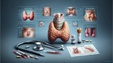
What an MRI shows
In addition to being safe, magnetic resonance imaging has another important advantage: its high diagnostic value. It produces high-quality images and allows for detailed examination of even the smallest structures. It is widely used to detect diseases of the thyroid gland, kidneys, liver, spine, spinal cord, joints, blood vessels, pancreas, etc. MRI is essential for diagnosis.
- Benign and malignant neoplasms, including metastases;
- Brain disorders;
- Musculoskeletal disorders;
- Biliary tract disorders, etc.
The diagnostic value of the examination can be enhanced by using contrast material. To do this, a contrast agent is administered intravenously to patients either prior to or during the procedure, which allows for the assessment of the neoplasm’s blood supply, examination of the pituitary gland, identification of demyelinating diseases, and determination of the tumor’s precise boundaries.
Indications and contraindications
The range of MRI applications is vast. For each group of conditions, a specific list of relevant diseases and conditions can be identified during the examination. For example, indications for MRI of the head and neck vessels include:

- Suspected aneurysm or malformation;
- Assessment of the degree of vascular stenosis;
- Hypertensive disorders;
- Diagnosis of congenital anomalies;
- For example, in cases of migraine headaches.
An MRI is not a unique or essential diagnostic procedure. There are many other tests that can help doctors make a diagnosis, assess the effectiveness of treatment, or detect certain conditions at an early stage. Decisions to include an MRI in a comprehensive diagnostic plan are always made on a case-by-case basis and only when necessary.
Like other imaging methods, MRI has a number of contraindications. These can be divided into several groups. The first group relates to the limitations of the MRI scanner itself. These limitations are usually related to the patient’s weight and waist circumference. Most MRI machines are designed for a maximum weight of 130–150 kg, but modern models allow for the examination of patients weighing 180 and even 200 kg. Additionally, in almost all cases, there is a restriction on maximum width—80 cm. The only option for such patients is an open MRI, where the examination is performed not in a tunnel but on a table (similar to an X-ray table).
")
The following categories include conditions under which an MRI scan is generally impossible. This group includes:
- Implanted electronic devices (e.g., pacemakers, insulin pumps, middle ear implants);
- The presence of hemostatic clamps made of ferromagnetic material;
- The presence of shrapnel or other metallic structures made of ferromagnetic materials in the patient’s body.
Finally, there are contraindications to MRI that apply only after special preparation or in cases of critical necessity. These contraindications include:
- Claustrophobia;
- Poorly compensated heart failure;
- Early pregnancy;
- Agitated patient;
- The need for constant monitoring of the patient’s condition, for example.
For example, if a patient is afraid of enclosed spaces or is agitated and unable to remain still in one position, sedation is prescribed before the MRI.
Preparation and procedure
In most cases, an MRI does not require special preparation. The exception is when examinations of the abdominal organs are performed. In these situations, you should avoid eating foods that cause gas the day before; on the day of the MRI, you should arrive at the clinic on an empty stomach. In all cases, you must remove any metal objects before the examination.
")
An MRI is performed while the patient is lying down. The duration of the scan depends largely on the size of the area being scanned. For example, an MRI of the brain takes about 15 minutes, while an MRI of the abdominal organs takes 40–45 minutes. During the scan, the patient must remain as still as possible; During an MRI, the patient should not feel any pain or other discomfort. The only discomfort is related to the relatively high noise level produced by the machine during operation. This issue can be addressed with special headphones.
After the examination is complete, the doctor interprets the images, makes a diagnosis, and prescribes treatment or further tests. If necessary, the data can be saved and photographed onto a removable storage device.